acromial extremity
flat, lateral end articulates with the acromion process of scapula forming the AC joint
S 2115 Anatomy & Physiology
Bone Markings of the Appendicular Skeleton
Objectives of the Appendicular Skeleton:
* To learn the names of the skeletal bones of the two girdles, shoulder and
pelvic and the limbs and their important landmarks.
* To understand the position on these landmarks associated with
articulating
surfaces and muscle attachments.
Using the bones in the lab, label each bone below with the markings listed in the right column:
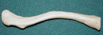
Right Clavicle (2) superior view
|
|
acromial extremity flat, lateral end articulates with the acromion process of scapula forming the AC joint |
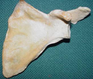
Left Scapula (2) anterior view
|
|
acromion process flaring projection at the lateral end, may be felt as tip of shoulder, articulates with clavicle glenoid cavity arm socket, articulates with head of humerus to form shoulder joint coracoid process hook-like projection, may be felt in groove between deltoid and pectoral muscles under clavicle |
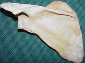
Left Scapula (2) posterior view
|
|
spine diagonal ridge along posterior surface ending as the acromion process
note: glenoid cavity and acromion process are visible |
Right Humerus anterior view Right Humerus posterior view
|
|
head smooth enlargement at proximal end, articulates with glenoid cavity surgical neck region below head, subject to fractures deltoid tuberosity V-shaped, rough area midway in the diaphysis, for insertion of deltoid muscle capitulum on distal epiphysis lateral knob for articulation with head of radius trochlea on distal epiphysis round medial projection for trochlear notch of ulna |
|
olecranon fossa depression on distal end for articulation with ulna
note: head and surgical neck are visible
|
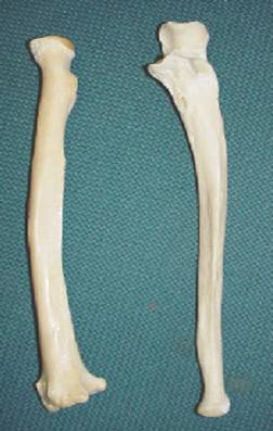
Right Radius and Ulna anterior view
|
|
RADIUS head of radius disc at proximal end of radius, articulates with capitulum of humerus and radial notch of ulna radial tuberosity medial projection below head for insertion of biceps muscles
ULNA trochlear notch c-curved notch which articulates with trochlea of humerus radial notch lateral depression inferior to trochlear notch, articulates with head of radius olecranon process note: not shown, your elbow, the blunt process on proximal end of ulna |
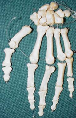
Hand anterior view
|
|
Carpals (8) proximal row from thumb side lunate (semilunar) articulates with radius triquetrum (triangular) pyramid shape pisiform pea-shaped distal row from thumb side trapezium trapezoid capitate hamate contains a hook-like process, with the pisiform the concave space forms the carpal tunnel
Metacarpals (5)
Phalanges (14 each hand) proximal phalanx articulates with metacarpals middle phalanx not in thumb distal phalanx |